XIAMEN GOHE MEDICAL TECHNOLOGY CO., LTD
Home

Blog

Blog

  • Understanding Orthopedic Internal Fixation Devices: Types, Materials & Applications
    Understanding Orthopedic Internal Fixation Devices: Types, Materials & Applications
    Apr 01, 2025
     Introduction  Orthopedic internal fixation devices are critical instruments in modern orthopedic trauma surgery. These devices are designed to stabilize fractured bones, playing a vital role in facilitating proper healing, reducing recovery time, and restoring function. In this article, we will review the various types of internal fixation devices, their materials, and clinical uses, helping healthcare professionals and distributors better understand the components of effective fracture management.    What Are Internal Fixation Devices?  Internal fixation refers to the surgical placement of devices within the body to stabilize broken bones. These implants hold fractured bone fragments in position, allowing the natural healing process to proceed with proper alignment and stability. Key benefits: Maintains bone alignment Promotes early mobilization Reduces the risk of nonunion or malunion    Common Types of Orthopedic Internal Fixation Devices  1. Plates and Screws These are the most commonly used devices, fixed directly onto the surface of the bone. Dynamic Compression Plates (DCP) Locking Compression Plates (LCP) Anatomical Plates Applications: Long bone fractures (humerus, femur, tibia), pelvic fractures, small bone fixation   2. Intramedullary Nails These metal rods are inserted into the marrow cavity of long bones. They provide excellent load-sharing properties and allow early weight-bearing after surgery. Applications: Femoral shaft, tibial shaft, and humeral shaft fractures Advantages: Minimally invasive, fast healing, early weight-bearing after surgery   3. Cable Systems These are used for complex fractures or bone reconstructions, particularly around joint areas. Material: Titanium alloy cables are commonly used for their flexibility and compatibility with bone tissue. Applications: Greater trochanter fractures, pelvic and acetabular reconstructions, spinal fixation.   4. Pedicle Screw Systems Part of spinal fixation systems, these screws are used to stabilize the vertebrae. Components: Pedicle screws, rods, transverse connectors Applications: Spinal trauma, deformity correction, spinal fusion surgeries    Materials Used in Internal Fixation Devices  Orthopedic implants must meet high material standards for strength, biocompatibility, and resistance to corrosion. The most commonly used materials are titanium alloy, stainless steel, and PEEK polymer. A comparison of these materials' properties is outlined below: Material Type Density (g/cm³) Young's Modulus (GPa) MRI Compatibility Corrosion Resistance Biocompatibility Strength (MPa) Characteristics Common Applications Titanium Alloy (Ti-6Al-4V) 4.43 110-120 Non-magnetic, Good High Excellent 900-1100 Lightweight, Corrosion-resistant, Excellent biocompatibility Bone plates, Screws, Intramedullary nails, Cable systems Stainless Steel (316L) 8.00 190-210 Magnetic, Affects scanning Moderate Good 520-600 High strength, Low cost, Good machinability Bone plates, Screws PEEK (Polyether Ether Ketone) 1.30 3.6 Non-magnetic, Good Excellent Excellent 90-120 Radiolucent, High flexibility Spinal fusion devices, Special replacement materials    Recovery & Post-Operative Care  Following surgery, patients typically undergo: Controlled rehabilitation Follow-up imaging (X-ray or CT scans) Monitoring for implant-related complications (e.g., loosening, infection) Optional implant removal 12–24 months after surgery (depending on the case)    Future Trends in Internal Fixatione  The field of orthopedic fixation is continuously advancing, with emerging technologies such as: 3D-printed custom implants Bioresorbable fixation materials Smart implants with integrated sensors Robot-assisted and navigated surgeries    Conclusion  Orthopedic internal fixation devices are fundamental to fracture treatment—ensuring stability, enhancing healing, and helping patients regain mobility more quickly. Whether you are a healthcare provider, distributor, or procurement professional, having a clear understanding of these devices is essential for improving orthopedic outcomes.  
    Read More
  • Disposable Puncture Suturing Device | Dual-Function Puncture and Suturing, Effortless Puncture, Perfect Closure
    Disposable Puncture Suturing Device | Dual-Function Puncture and Suturing, Effortless Puncture, Perfect Closure
    Feb 11, 2025
    Laparoscopic surgery, as a minimally invasive technique, has been widely adopted in general surgery for various procedures. During laparoscopic surgery, surgical instruments access the abdominal cavity through trocars. After the procedure, puncture sites remain on the abdominal wall. Improper suturing of these sites can lead to complications such as bleeding, hematoma, infection, and trocar site herniation. Suturing the fascial layer of the puncture site effectively reduces the difficulty of incision closure in laparoscopic surgery, minimizes the risk of trocar site hernia, and enhances surgical safety. Trocars provide access for endoscopic instruments during laparoscopic surgery. A trocar with integrated suturing capability combines fascial closure functionality into the device, enabling both puncture and suturing in a single step. The disposable puncture suturing device consists of a cannula with a guide hole, a visual obturator, a suturing puncture needle with a side hole, and an abdominal wall puncture needle clamp for grasping the suture. The guide hole on the cannula is sealed with an ultra-thin silicone membrane to prevent air leakage during use. ▲ Schematic Diagram of Fascial Closure Mechanism   Product Advantages Combines dual functions of puncture and suturing, eliminating the need for a separate fascial closure device—offering both medical and economic value. • Precise Visual Puncture: Significantly reduces the risk of organ injury. • Simplified Workflow: Easy to master with minimal training, ensuring efficient operation. • Time-Saving Technology: Rapid suturing enhances procedural efficiency. • Clear Visual Suturing: The suturing process is fully visible, ensuring accuracy and avoiding organ damage. • Broad Compatibility: The obturator is compatible with various commercially available cannulas—bladed or bladeless, disposable or reusable.   Strong Market Demand Data indicates a substantial and steadily growing market demand for laparoscopic trocars. In 2019, sales of disposable trocars in China reached 18.4 million units, with a market value of RMB 1.77 billion. By 2024, this is projected to grow to RMB 5.091 billion, reflecting a compound annual growth rate (CAGR) of 23.4%. Amid the trend of declining prices due to centralized medical procurement, conventional laparoscopic trocars are losing their competitive edge. In contrast, laparoscopic trocars with fascial closure functionality demonstrate significant clinical advantages. Their innovative design integrates both puncture access and fascial closure, aligning with the market's demand for advanced medical devices.
    Read More
  • Subtalar Arthroereisis for the Treatment of Flatfoot
    Subtalar Arthroereisis for the Treatment of Flatfoot
    Nov 01, 2024
    What is Flatfoot? Flatfoot, as the name suggests, refers to the collapse of the arch of the foot. In fact, flatfoot is a multi-dimensional deformity, not only manifested as the collapse of the medial longitudinal arch but also including rearfoot valgus, and in severe cases, it may even cause ankle valgus. The collapse of the arch and rearfoot valgus are often accompanied by a pronation deformity of the forefoot. Collapse of the Arch in Flatfoot   Rearfoot Valgus and Medial Prominence in Flatfoot   Do Flatfeet Need Treatment and When? Generally speaking, the incidence of flatfoot is high in children under the age of 10, but as growth and development progress, the arch of the foot will gradually improve and recover. If after the age of 10, the arch has not recovered, it should be taken seriously. If there are long-term symptoms such as pain, and the pain cannot be relieved even with the use of arch supports, or if the degree of deformity is severe; or if there is limited joint mobility, and the arch cannot be passively corrected when checked, i.e., so-called rigid flatfoot, then it is necessary to seek medical attention.   What is Subtalar Joint Bracing Surgery? Subtalar joint bracing surgery is a surgical intervention measure for the further treatment of flatfoot that is unresponsive to conservative treatment. It is a minimally invasive treatment method that corrects the deformity of flatfoot by placing a "screw" between the sinus tarsi, talus, and calcaneus, known as a subtalar joint stabilization screw. Correction of Talus Internal Rotation After Bracing   Correction of Talus Sinking After Bracing   Recovery of the Arch After Bracing Surgery   Correction of Rearfoot Valgus After Bracing Surgery   Postoperative Recovery After the placement of the brace, one can walk as soon as the wound heals, which generally takes about two weeks. To avoid long-term impact on the joint from the device placed inside the joint, it is recommended to remove the subtalar joint stabilization screw after one year.
    Read More
  • Efficient Orthodontic Assistant——Anchorage Mini-Implant
    Efficient Orthodontic Assistant——Anchorage Mini-Implant
    Aug 18, 2024
     What is an anchorage mini-implant?  The anchorage mini-implant, also known as a micro-screw implant or bone anchor, can be simply understood as a means or device that serves as a fixed point for orthodontic forces. In conventional orthodontic treatments, such as wearing braces, the fulcrum for orthodontic forces is provided by other more robust teeth, which generate traction to move the teeth that need to be shifted to their designated positions. This process is akin to a tug-of-war; without a significant difference in strength, it's generally a situation of you advance and I retreat, I advance and you retreat, a contest of tenacity and endurance. Even the most robust teeth can be at risk of being dragged forward, which is, of course, a major taboo in orthodontic processes. Perfection-seeking dentists absolutely cannot tolerate such situations that affect the effect of teeth retraction. The emergence of anchorage mini-implants has effectively addressed the aforementioned issues. They can be fixed within the bone tissue, providing absolute anchorage. When applying traction to misaligned teeth, they can better control the movement of the teeth, in which direction they should move, and how much they should move.    Do all orthodontic treatments require anchorage mini-implants?  Of course not. Anchorage mini-implants are particularly effective for patients with severe skeletal deformities. If your correction involves the following situations, then the power of bone anchors will be needed. 1. Patients with protruding teeth: Those who need to improve the facial protrusion issue. By placing anchorage mini-implants, the protruding front teeth can be retracted, thereby maximizing the improvement of protrusion and improving the side profile. 2. Lowering upper and lower front teeth: With the help of anchorage mini-implants between the roots of the upper and lower front teeth, conditions such as uncoordinated lip-tooth relationships, gummy smiles, deep overbites, and underbites, caused by excessive eruption of upper and lower front teeth, can be improved by directly applying pressure to the anterior archwire through a chain loop, which is a simple and effective method. 3. When there are frequent issues with posterior teeth: When a posterior tooth is missing on one side, making it difficult to control the midline, anchorage mini-implants can be used; when posterior teeth are obstructing growth, anchorage mini-implants can also be used to straighten the tilted posterior teeth. Many patients have insufficient understanding of anchorage mini-implants and have many doubts. Now let's address the common questions to alleviate your fears about getting anchorage mini-implants:    9 Questions About Anchorage Mini-Implants  1. What is the use of anchorage mini-implants? Tooth movement is the result of the interaction between force and counterforce. The role of anchorage mini-implants is to resist the counterforce in place of teeth, providing a stable force for the teeth that need to be moved while avoiding unnecessary movement of other teeth. 2. What material are anchorage mini-implants made of? Can they cause allergies? Anchorage mini-implants are generally made of pure titanium, titanium alloy, or stainless steel. They are required to have good biocompatibility and sufficient hardness to prevent the bone anchor from breaking during insertion. Before entering clinical use, they must first undergo biocompatibility testing and then clinical trials. Therefore, they do not cause allergic reactions. 3. Are there any risks with anchorage mini-implants? The procedure for placing anchorage mini-implants generally involves disinfection, local anesthesia injection, and finally, the implantation of the mini-implant. The anesthesia is a local anesthetic, so there is no risk involved. 4. Does getting an anchorage mini-implant hurt? Local anesthesia is required before placing an anchorage mini-implant, so there is no pain. After the effect of the anesthetic wears off, there may be local pain and discomfort. If the pain is severe, you can take painkillers as prescribed by a doctor, and it usually takes about three days to fully recover. 
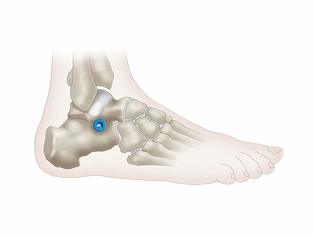
5. Where are anchorage mini-implants usually placed? The placement of anchorage mini-implants needs to be coordinated with individual correction plans and varies from person to person. Whether it's the posterior or anterior tooth area, they are usually implanted between two teeth, avoiding the tooth roots. Special locations may also be chosen according to actual needs, such as the external oblique line of the mandible. 6. How should anchorage mini-implants be cared for? When brushing your teeth, you can gently brush the anchorage mini-implant a few times to clean the food debris around it, or you can use a water flosser to rinse it. However, it is important not to use a vibrating electric toothbrush to clean the anchorage mini-implant to avoid loosening it. 7. Can anchorage mini-implants become loose? What if they do? Anchorage mini-implants can become loose, for example, if food debris around the anchorage mini-implant is not cleaned, causing inflammation, the anchorage mini-implant will become loose. If it becomes loose, the anchorage mini-implant needs to be removed, disinfected, and replaced in a new position or re-implanted after the alveolar bone has recovered. 8. When should anchorage mini-implants be removed? When anchorage mini-implants are no longer needed, they can be removed. 9. Will there be a wound after the anchorage mini-implant is removed? Yes, there will be, but it will usually heal automatically within three to five days, so there is no need to worry.
    Read More

Leave a message

Leave a message
If you are interested in our products and want to know more details, please leave a message here, we will reply you as soon as we can.
submit
Contact Us:info@gohemed.com

Home

Products

whatsApp

contact